Arm Muscles Diagram Posterior - Muscles Of Right Upper Arm Photograph by Asklepios Medical ... / Arm anatomy diagram for artists.
byAdmin•
0
Arm Muscles Diagram Posterior - Muscles Of Right Upper Arm Photograph by Asklepios Medical ... / Arm anatomy diagram for artists.. Learn vocabulary, terms and more with flashcards, games and other study tools. Abducts the ulna when the arm is pronating (rotating forearm medially), helps triceps brachii extend forearm b. The anterior and the posterior compartments of the arm. The anterior compartment is the flexor compartment because these we've just got a diagram of it here. The extensor muscles can be individually visible on a flexed arm, even on non muscular people.
Arm muscle diagram shoulder and arm muscles posterior diagram quizlet. Arm muscle diagram muscles of the rotator cuff human anatomy and physiology lab bsb 141. The radial nerve is the largest branch of brachial plexus travels along with. Only two of these do not originate on the scapula, the pectoralis major and the latissumus extensor digitorum. Nine muscles cross the shoulder joint.
Shoulder Muscles Diagram / Applied Anatomy Of The Shoulder ... from o.quizlet.com Draw labelled diagram showing branches of profunda brachi artery. Two intermuscular septa (medial and lateral) extend from it to attach to the humerus at the medial condylar ridge and lateral supracondylar ridge, respectively. From lateral epicondyle of the humerus d. From the arm muscle diagram above, the muscles of the arm that can be seen easily on the surface include biceps, triceps, brachioradialis, extensor carpi radialis longus, and deltoid. The anterior compartment is the flexor compartment because these we've just got a diagram of it here. The muscles of the upper arm are responsible for the flexion and extension of the forearm at the elbow joint. Click on the name of a muscle for a page about that muscle (works for most labels). Forearm muscles anatomy, posterior arm muscles, muscles of the arm and forearm, forearm anatomy, arm muscles diagram, deep muscles of forearm, muscles in lower arm.
For more anatomy content please follow us and visit our website:
Flexion of the forearm is achieved by a group of three on the posterior side of the arm the extensor muscles, such as the extensor carpi ulnaris and extensor digitorum, act as antagonists to. A sheath of deep fascia surrounds the arm, the brachial fascia. Arm muscle diagram muscles of the rotator cuff human anatomy and physiology lab bsb 141. Tutorial covers the muscles of the anterior and posterior compartment of the upper arm, and talks about nerve supply and actions of these muscles. Muscles that move the arm. Lateral surface of the olecranon process of ulna and the superior proximal part of the posterior ulna. The arm muscles comprise five muscles, which. Learn the muscles of the arm with free quizzes, diagrams and worksheets. Related posts of shoulder muscles labelled diagram. Tutorials and quizzes on muscles that act on the arm/humerus (arm muscles: Two intermuscular septa (medial and lateral) extend from it to attach to the humerus at the medial condylar ridge and lateral supracondylar ridge, respectively. On the posterior side of the arm are the extensor muscles, such as the extensor carpi ulnaris, and extensor digitorum which act as antagonist to the flexor muscles by extending the hand and fingers. (redirected from posterior compartment of the arm).
The fascial compartments of arm refers to the specific anatomical term of the compartments within the upper segment of the upper limb(the arm) of the body. The radial nerve is the largest branch of brachial plexus travels along with. Only two of these do not originate on the scapula, the pectoralis major and the latissumus extensor digitorum. There are anterior muscles diagrams and posterior muscles diagrams. Lower left arm, posterior view, back of hand facing front.
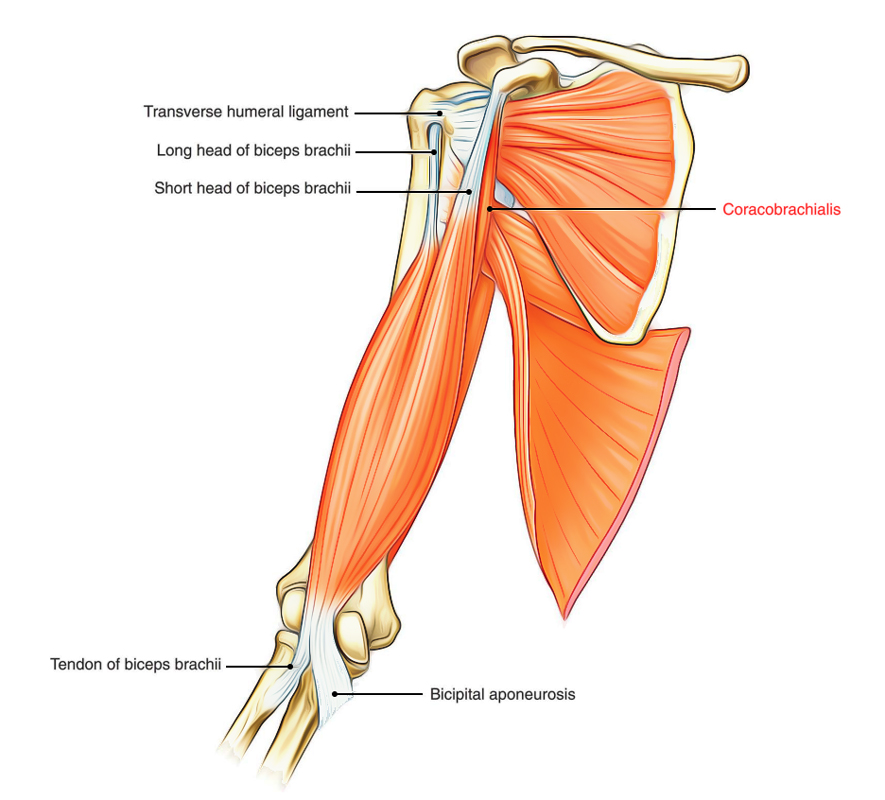
muscles of the upper limb, front or anterior view | Muscle ... from i.pinimg.com The delta, it can rolls back, especially the further back. Posterior muscles of the arm and forearm. There are anterior muscles diagrams and posterior muscles diagrams. Coracobrachialis brachialis biceps brachii coracobrachialis: Want to learn more about it? Whole arm muscles posterior labeled stock illustration 155445635 these pictures of this page are about:deep posterior arm muscles. Tutorial covers the muscles of the anterior and posterior compartment of the upper arm, and talks about nerve supply and actions of these muscles. Anterior arm muscle diagram anterior forearm deep anatomy gray s illustration radiology case.
Related posts of shoulder muscles labelled posterior muscles of the body diagram (with images).
We hope this picture posterior view of human body muscles diagram can help you study and research. Whole arm muscles posterior labeled stock illustration 155445635 these pictures of this page are about:deep posterior arm muscles. Nine muscles cross the shoulder joint. Study flashcards on muscles of the posterior arm at cram.com. The sacrum bone is almost always noticeable, no matter what the body type, because it is not covered with muscles or substantial fatty tissue. There are anterior muscles diagrams and posterior muscles diagrams. Two intermuscular septa (medial and lateral) extend from it to attach to the humerus at the medial condylar ridge and lateral supracondylar ridge, respectively. Coracobrachialis brachialis biceps brachii coracobrachialis: Arm anatomy diagram for artists. Flexion of the forearm is achieved by a group of three on the posterior side of the arm the extensor muscles, such as the extensor carpi ulnaris and extensor digitorum, act as antagonists to. Arm muscle diagram shoulder and arm muscles posterior diagram quizlet. Learn vocabulary, terms and more with flashcards, games and other study tools. The muscles of the upper arm are split into anterior and posterior compartments.
The extensor muscles can be individually visible on a flexed arm, even on non muscular people. From the arm muscle diagram above, the muscles of the arm that can be seen easily on the surface include biceps, triceps, brachioradialis, extensor carpi radialis longus, and deltoid. Draw labelled diagram showing branches of profunda brachi artery. Tutorials and quizzes on muscles that act on the arm/humerus (arm muscles: From lateral epicondyle of the humerus d.
Easy Notes On 【Muscles of the Upper Arm】Learn in Just 3 ... from www.earthslab.com On the posterior side of the arm are the extensor muscles, such as the extensor carpi ulnaris, and extensor digitorum which act as antagonist to the flexor muscles by extending the hand and fingers. Arm muscle diagram muscles of the rotator cuff human anatomy and physiology lab bsb 141. Lateral surface of the olecranon process of ulna and the superior proximal part of the posterior ulna. Learn the muscles of the arm with free quizzes, diagrams and worksheets. Muscles that cross the elbow (moving the forearm) (posterior view). Learn vocabulary, terms and more with flashcards, games and other study tools. Whole arm muscles posterior labeled stock illustration 155445635 these pictures of this page are about:deep posterior arm muscles. From lateral epicondyle of the humerus d.
For the start postion, lift your arms in front of you.
For more anatomy content please follow us and visit our website: Muscles of anterior (flexor) compartment of arm, their origin, insertion, action/s and nerve supply are as follows superior ulnar collateral branch of brachial artery. On the posterior side of the arm are the extensor muscles, such as the extensor carpi ulnaris, and extensor digitorum which act as antagonist to the flexor muscles by extending the hand and fingers. This is the coracoid process. Extension at joints of fingers and wrist. Arm anatomy diagram for artists. For the start postion, lift your arms in front of you. Two intermuscular septa (medial and lateral) extend from it to attach to the humerus at the medial condylar ridge and lateral supracondylar ridge, respectively. Muscles flexors in the arm all innervated the musculocutaneous nerve: Draw labelled diagram showing branches of profunda brachi artery. Learn vocabulary, terms and more with flashcards, games and other study tools. Related posts of shoulder muscles labelled diagram. The muscles of the upper arm are responsible for the flexion and extension of the forearm at the.
The radial nerve is the largest branch of brachial plexus travels along with arm muscles diagram. Learn more about their anatomy at kenhub!